Figure 3
Angioarchitectonics of acute pneumonia
Klepikov Igor*
Published: 07 February, 2019 | Volume 4 - Issue 1 | Pages: 018-022
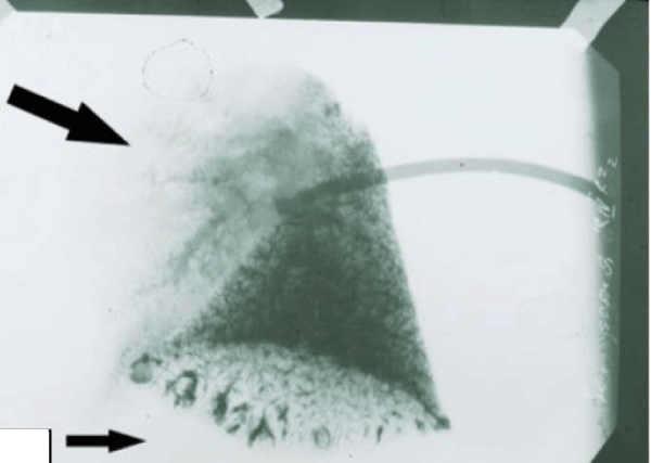
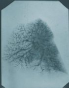
Figure 3:
Venogram of the lung with total inflammation of the upper lobe. (Subsequent anatomical and histological studies revealed no necrotic changes). A sharp depletion of the venous pattern in the upper affected part of the lung(big arrow). In the lower part of the architectonics of the vessels is preserved, but against this background, relatively large vascular formations of semicircular shape are revealed(little arrow), which can be a reflection of overload and compensatory blood flow.
Read Full Article HTML DOI: 10.29328/journal.jcicm.1001020 Cite this Article Read Full Article PDF
More Images
Similar Articles
-
Evaluation of Desmin, α-SMA and hTERT expression in pulmonary fibrosis and lung cancerFarahnaz Fallahian*,Seyed Ali Javad Moosavi,Frouzandeh Mahjoubi,Samira Shabani,Pegah Babaheidarian,Tayebeh Majidzadeh. Evaluation of Desmin, α-SMA and hTERT expression in pulmonary fibrosis and lung cancer. . 2018 doi: 10.29328/journal.jcicm.1001011; 3: 001-009
-
Dependence of the results of treatment of acute pneumonia on the doctrine of the diseaseIgor Klepikov*. Dependence of the results of treatment of acute pneumonia on the doctrine of the disease. . 2018 doi: 10.29328/journal.jcicm.1001012; 3: 010-012
-
Acute pneumonia: Facts and realities against etiological hypotheses and beliefsKlepikov Igor*. Acute pneumonia: Facts and realities against etiological hypotheses and beliefs. . 2019 doi: 10.29328/journal.jcicm.1001019; 4: 010-017
-
Angioarchitectonics of acute pneumoniaKlepikov Igor*. Angioarchitectonics of acute pneumonia. . 2019 doi: 10.29328/journal.jcicm.1001020; 4: 018-022
-
Do you really want to improve the results of treatment for acute pneumonia?Klepikov Igor*. Do you really want to improve the results of treatment for acute pneumonia?. . 2019 doi: 10.29328/journal.jcicm.1001021; 4: 023-027
-
Prognosis factors for dengue shock syndrome in childrenEka Fitri Sari Ningrum*. Prognosis factors for dengue shock syndrome in children. . 2021 doi: 10.29328/journal.jcicm.1001039; 6: 033-037
-
Clinical profile, etiology, outcome and new-onset diabetes: A SARI case seriesSiddharth Agarwal*,Sapna Agarwal,Raj Kumar Verma,Shreyash Dayal. Clinical profile, etiology, outcome and new-onset diabetes: A SARI case series. . 2022 doi: 10.29328/journal.jcicm.1001041; 7: 005-015
-
Why is Pain not Characteristic of Inflammation of the Lung Tissue?Igor Klepikov*. Why is Pain not Characteristic of Inflammation of the Lung Tissue?. . 2024 doi: 10.29328/journal.jcicm.1001046; 9: 005-007
Recently Viewed
-
Evaluation of Heavy Metals in Commercial Baby FoodsOmobolanle David Garuba, Judith C Anglin, Sonya Good, Shodimu-Emmanuel Olufemi, Olubukola Monisola Oyawoye, Sodipe Ayodotun*. Evaluation of Heavy Metals in Commercial Baby Foods. Arch Food Nutr Sci. 2024: doi: 10.29328/journal.afns.1001056; 8: 012-020
-
Endoscopic treatment of pancreatic diseases via Duodenal Minor Papilla: 135 cases treated by Sphincterotomy, Endoscopic Pancreatic Duct Balloon Dilation (EPDBD), and Pancreatic Stenting (EPS)Tadao Tsuji*, G Sun,A Sugiyama, Y Amano,S Mano,T Shinobi,H Tanaka,M Kubochi,K Ohishi,Y Moriya,M Ono,T Masuda, H Shinozaki,H Kaneda,H Katsura,T Mizutani, K Miura,M Katoh, K Yamafuji, K Takeshima,N Okamoto,Y Hoshino,N Tsurumi,S Hisada,J Won,T Kogiso,K Yatsuji,M Iimura, T Kakimoto,S Nyuhzuki. Endoscopic treatment of pancreatic diseases via Duodenal Minor Papilla: 135 cases treated by Sphincterotomy, Endoscopic Pancreatic Duct Balloon Dilation (EPDBD), and Pancreatic Stenting (EPS) . Ann Clin Gastroenterol Hepatol. 2019: doi: 10.29328/journal.acgh.1001009; 3: 012-019
-
Euthanasia: Growing Acceptance amid Lingering ReluctanceTshibambe N Tshimbombu,Immanuel Olarinde,Judea Wiggins*,Maxwell Vergo. Euthanasia: Growing Acceptance amid Lingering Reluctance. Clin J Nurs Care Pract. 2025: doi: 10.29328/journal.cjncp.1001058; 9: 001-006
-
Various Theories of Fast and Ultrafast Magnetization DynamicsManfred Fähnle*. Various Theories of Fast and Ultrafast Magnetization Dynamics. Int J Phys Res Appl. 2024: doi: 10.29328/journal.ijpra.1001101; 7: 154-158
-
Nitrogen Fixation and Yield of Common Bean Varieties in Response to Shade and Inoculation of Common BeanSelamawit Assegid*,Girma Abera. Nitrogen Fixation and Yield of Common Bean Varieties in Response to Shade and Inoculation of Common Bean. J Plant Sci Phytopathol. 2023: doi: 10.29328/journal.jpsp.1001122; 7: 157-162
Most Viewed
-
Evaluation of Biostimulants Based on Recovered Protein Hydrolysates from Animal By-products as Plant Growth EnhancersH Pérez-Aguilar*, M Lacruz-Asaro, F Arán-Ais. Evaluation of Biostimulants Based on Recovered Protein Hydrolysates from Animal By-products as Plant Growth Enhancers. J Plant Sci Phytopathol. 2023 doi: 10.29328/journal.jpsp.1001104; 7: 042-047
-
Sinonasal Myxoma Extending into the Orbit in a 4-Year Old: A Case PresentationJulian A Purrinos*, Ramzi Younis. Sinonasal Myxoma Extending into the Orbit in a 4-Year Old: A Case Presentation. Arch Case Rep. 2024 doi: 10.29328/journal.acr.1001099; 8: 075-077
-
Feasibility study of magnetic sensing for detecting single-neuron action potentialsDenis Tonini,Kai Wu,Renata Saha,Jian-Ping Wang*. Feasibility study of magnetic sensing for detecting single-neuron action potentials. Ann Biomed Sci Eng. 2022 doi: 10.29328/journal.abse.1001018; 6: 019-029
-
Pediatric Dysgerminoma: Unveiling a Rare Ovarian TumorFaten Limaiem*, Khalil Saffar, Ahmed Halouani. Pediatric Dysgerminoma: Unveiling a Rare Ovarian Tumor. Arch Case Rep. 2024 doi: 10.29328/journal.acr.1001087; 8: 010-013
-
Physical activity can change the physiological and psychological circumstances during COVID-19 pandemic: A narrative reviewKhashayar Maroufi*. Physical activity can change the physiological and psychological circumstances during COVID-19 pandemic: A narrative review. J Sports Med Ther. 2021 doi: 10.29328/journal.jsmt.1001051; 6: 001-007

HSPI: We're glad you're here. Please click "create a new Query" if you are a new visitor to our website and need further information from us.
If you are already a member of our network and need to keep track of any developments regarding a question you have already submitted, click "take me to my Query."


