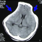
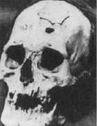
Figure 4
Sinking Skin Flap Syndrome
Liew BS*, Rosman AK and Adnan JS
Published: 08 September, 2017 | Volume 2 - Issue 1 | Pages: 042-048
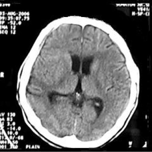
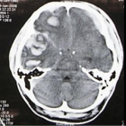
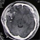
Figure 4:
Computed tomography (CT) scan brain (axial view) showing no extraaxial collection, no enchancing lesion, no midline-shift, with encephalomalacia over right temporoparietal area. The sulci and gyri are well-visualised without any effacement.
Read Full Article HTML DOI: 10.29328/journal.jcicm.1001009 Cite this Article Read Full Article PDF
More Images
Similar Articles
-
Phenibut Overdose in Combination with Fasoracetam: Emerging Drugs of AbuseCristian Merchan*,Ryan Morgan,John Papadopoulos,David Fridman. Phenibut Overdose in Combination with Fasoracetam: Emerging Drugs of Abuse. . 2016 doi: 10.29328/journal.jcicm.1001001; 1: 001-004
-
Nursing Care of ICU Patients Lightly Sedated with DexmedetomidineÅsa Engström*,Maria Johansson,Mia Mattsson,Ulrica Strömbäck. Nursing Care of ICU Patients Lightly Sedated with Dexmedetomidine. . 2016 doi: 10.29328/journal.jcicm.1001002; 1: 005-013
-
Complicated Hepatitis A Virus Infection: A Report of Three Cases from Single Tertiary Referral CenterOmkolsoum M Alhaddad,Maha M Elsabaawy*,Khalid A Gameel,Marwa Elfauomy,Olfat Hendy,Eman A Rewisha. Complicated Hepatitis A Virus Infection: A Report of Three Cases from Single Tertiary Referral Center. . 2016 doi: 10.29328/journal.jcicm.1001003; 1: 014-020
-
Critical Management of Status EpilepticusFarahnaz Fallahian*,Seyed MohammadReza Hashemian*. Critical Management of Status Epilepticus. . 2017 doi: 10.29328/journal.jcicm.1001004; 2: 001-015
-
Comparative Hemodynamic Evaluation of the LUCAS® Device and Manual Chest Compression in Patients with Out-of-Hospital Cardiac ArrestMirek S,Opprecht N*,Daisey A,Milojevitch E,Soudry- Faure A,Freysz M. Comparative Hemodynamic Evaluation of the LUCAS® Device and Manual Chest Compression in Patients with Out-of-Hospital Cardiac Arrest. . 2017 doi: 10.29328/journal.jcicm.1001005; 2: 016-024
-
Chemotherapy Exposure and outcomes of Chronic Lymphoid Leukemia PatientsJosephina G Kuiper*,Patience Musingarimi,Christoph Tapprich,Fernie JA Penning-van Beest,Maren Gaudig. Chemotherapy Exposure and outcomes of Chronic Lymphoid Leukemia Patients. . 2017 doi: 10.29328/journal.jcicm.1001006; 2: 025-033
-
Knowledge, attitude and practices associated with diagnosis and management of Skin and Soft Tissue Infections (SSTIs) among Pediatric Residents and Physicians in a Tertiary Hospital in United Arab Emirates (UAE)Eiman Al Blooshi,Farah Othman,Abeer Al Naqbi,Majid Al Rumaithi,Khawla Fikry,Mariam Al Jneibi,Hossam Al Tatari*. Knowledge, attitude and practices associated with diagnosis and management of Skin and Soft Tissue Infections (SSTIs) among Pediatric Residents and Physicians in a Tertiary Hospital in United Arab Emirates (UAE) . . 2017 doi: 10.29328/journal.jcicm.1001007; 2: 034-039
-
Unusual presentation of a bilateral basilar stroke: BradycardiaZidouh S*,Jidane S,Nabhani T,Chouaib N,Sirbou R,Belkouch,Belyamani L. Unusual presentation of a bilateral basilar stroke: Bradycardia. . 2017 doi: 10.29328/journal.jcicm.1001008; 2: 040-041
-
Sinking Skin Flap SyndromeLiew BS*,Rosman AK,Adnan JS. Sinking Skin Flap Syndrome. . 2017 doi: 10.29328/journal.jcicm.1001009; 2: 042-048
-
Intensive Care Units (ICU): The clinical pharmacist role to improve clinical outcomes and reduce mortality rate- An undeniable functionLuisetto M*,Ghulam Rasool Mashori. Intensive Care Units (ICU): The clinical pharmacist role to improve clinical outcomes and reduce mortality rate- An undeniable function. . 2017 doi: 10.29328/journal.jcicm.1001010; 2: 049-056
Recently Viewed
-
Obesity in Patients with Chronic Obstructive Pulmonary Disease as a Separate Clinical PhenotypeDaria A Prokonich*, Tatiana V Saprina, Ekaterina B Bukreeva. Obesity in Patients with Chronic Obstructive Pulmonary Disease as a Separate Clinical Phenotype. J Pulmonol Respir Res. 2024: doi: 10.29328/journal.jprr.1001060; 8: 053-055
-
Scientific Analysis of Eucharistic Miracles: Importance of a Standardization in EvaluationKelly Kearse*,Frank Ligaj. Scientific Analysis of Eucharistic Miracles: Importance of a Standardization in Evaluation. J Forensic Sci Res. 2024: doi: 10.29328/journal.jfsr.1001068; 8: 078-088
-
Evolution of the Mineralocorticoid Receptor and Gender Difference in Cardiovascular PathologyAlessandro Zuccalà*. Evolution of the Mineralocorticoid Receptor and Gender Difference in Cardiovascular Pathology. J Cardiol Cardiovasc Med. 2025: doi: 10.29328/journal.jccm.1001204; 10: 008-015
-
Clinical and immunological characteristics of depressive patients with a clinical high risk of schizophreniaOmelchenko MA,Zozulya SA,Kaleda VG,Klyushnik TP*. Clinical and immunological characteristics of depressive patients with a clinical high risk of schizophrenia. Insights Depress Anxiety. 2023: doi: 10.29328/journal.ida.1001034; 7: 001-003
-
Atypical Anti-GBM with ANCA Vasculitis- A Rarest of the Rare Entity: Index Case from Eastern IndiaGopambuj Singh Rathod*, Atanu Pal, Pallavi Mahato, Aakash Roy, Debroop Sengupta, Muzzamil Ahmad. Atypical Anti-GBM with ANCA Vasculitis- A Rarest of the Rare Entity: Index Case from Eastern India. J Clini Nephrol. 2024: doi: 10.29328/journal.jcn.1001139; 8: 124-126
Most Viewed
-
Evaluation of Biostimulants Based on Recovered Protein Hydrolysates from Animal By-products as Plant Growth EnhancersH Pérez-Aguilar*, M Lacruz-Asaro, F Arán-Ais. Evaluation of Biostimulants Based on Recovered Protein Hydrolysates from Animal By-products as Plant Growth Enhancers. J Plant Sci Phytopathol. 2023 doi: 10.29328/journal.jpsp.1001104; 7: 042-047
-
Sinonasal Myxoma Extending into the Orbit in a 4-Year Old: A Case PresentationJulian A Purrinos*, Ramzi Younis. Sinonasal Myxoma Extending into the Orbit in a 4-Year Old: A Case Presentation. Arch Case Rep. 2024 doi: 10.29328/journal.acr.1001099; 8: 075-077
-
Feasibility study of magnetic sensing for detecting single-neuron action potentialsDenis Tonini,Kai Wu,Renata Saha,Jian-Ping Wang*. Feasibility study of magnetic sensing for detecting single-neuron action potentials. Ann Biomed Sci Eng. 2022 doi: 10.29328/journal.abse.1001018; 6: 019-029
-
Pediatric Dysgerminoma: Unveiling a Rare Ovarian TumorFaten Limaiem*, Khalil Saffar, Ahmed Halouani. Pediatric Dysgerminoma: Unveiling a Rare Ovarian Tumor. Arch Case Rep. 2024 doi: 10.29328/journal.acr.1001087; 8: 010-013
-
Physical activity can change the physiological and psychological circumstances during COVID-19 pandemic: A narrative reviewKhashayar Maroufi*. Physical activity can change the physiological and psychological circumstances during COVID-19 pandemic: A narrative review. J Sports Med Ther. 2021 doi: 10.29328/journal.jsmt.1001051; 6: 001-007

HSPI: We're glad you're here. Please click "create a new Query" if you are a new visitor to our website and need further information from us.
If you are already a member of our network and need to keep track of any developments regarding a question you have already submitted, click "take me to my Query."


