Figure 3
Sinking Skin Flap Syndrome
Liew BS*, Rosman AK and Adnan JS
Published: 08 September, 2017 | Volume 2 - Issue 1 | Pages: 042-048
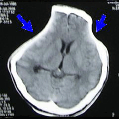
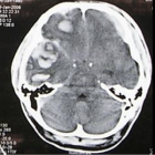
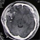
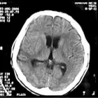
Figure 3:
Computed tomography (CT) scan brain (axial view) showing sinking skin flap bilaterally with compression over cerebral hemispheres bilaterally with effacement of sulci and gyri (marked by two arrows).
Read Full Article HTML DOI: 10.29328/journal.jcicm.1001009 Cite this Article Read Full Article PDF
More Images
Similar Articles
-
Phenibut Overdose in Combination with Fasoracetam: Emerging Drugs of AbuseCristian Merchan*,Ryan Morgan,John Papadopoulos,David Fridman. Phenibut Overdose in Combination with Fasoracetam: Emerging Drugs of Abuse. . 2016 doi: 10.29328/journal.jcicm.1001001; 1: 001-004
-
Nursing Care of ICU Patients Lightly Sedated with DexmedetomidineÅsa Engström*,Maria Johansson,Mia Mattsson,Ulrica Strömbäck. Nursing Care of ICU Patients Lightly Sedated with Dexmedetomidine. . 2016 doi: 10.29328/journal.jcicm.1001002; 1: 005-013
-
Complicated Hepatitis A Virus Infection: A Report of Three Cases from Single Tertiary Referral CenterOmkolsoum M Alhaddad,Maha M Elsabaawy*,Khalid A Gameel,Marwa Elfauomy,Olfat Hendy,Eman A Rewisha. Complicated Hepatitis A Virus Infection: A Report of Three Cases from Single Tertiary Referral Center. . 2016 doi: 10.29328/journal.jcicm.1001003; 1: 014-020
-
Critical Management of Status EpilepticusFarahnaz Fallahian*,Seyed MohammadReza Hashemian*. Critical Management of Status Epilepticus. . 2017 doi: 10.29328/journal.jcicm.1001004; 2: 001-015
-
Comparative Hemodynamic Evaluation of the LUCAS® Device and Manual Chest Compression in Patients with Out-of-Hospital Cardiac ArrestMirek S,Opprecht N*,Daisey A,Milojevitch E,Soudry- Faure A,Freysz M. Comparative Hemodynamic Evaluation of the LUCAS® Device and Manual Chest Compression in Patients with Out-of-Hospital Cardiac Arrest. . 2017 doi: 10.29328/journal.jcicm.1001005; 2: 016-024
-
Chemotherapy Exposure and outcomes of Chronic Lymphoid Leukemia PatientsJosephina G Kuiper*,Patience Musingarimi,Christoph Tapprich,Fernie JA Penning-van Beest,Maren Gaudig. Chemotherapy Exposure and outcomes of Chronic Lymphoid Leukemia Patients. . 2017 doi: 10.29328/journal.jcicm.1001006; 2: 025-033
-
Knowledge, attitude and practices associated with diagnosis and management of Skin and Soft Tissue Infections (SSTIs) among Pediatric Residents and Physicians in a Tertiary Hospital in United Arab Emirates (UAE)Eiman Al Blooshi,Farah Othman,Abeer Al Naqbi,Majid Al Rumaithi,Khawla Fikry,Mariam Al Jneibi,Hossam Al Tatari*. Knowledge, attitude and practices associated with diagnosis and management of Skin and Soft Tissue Infections (SSTIs) among Pediatric Residents and Physicians in a Tertiary Hospital in United Arab Emirates (UAE) . . 2017 doi: 10.29328/journal.jcicm.1001007; 2: 034-039
-
Unusual presentation of a bilateral basilar stroke: BradycardiaZidouh S*,Jidane S,Nabhani T,Chouaib N,Sirbou R,Belkouch,Belyamani L. Unusual presentation of a bilateral basilar stroke: Bradycardia. . 2017 doi: 10.29328/journal.jcicm.1001008; 2: 040-041
-
Sinking Skin Flap SyndromeLiew BS*,Rosman AK,Adnan JS. Sinking Skin Flap Syndrome. . 2017 doi: 10.29328/journal.jcicm.1001009; 2: 042-048
-
Intensive Care Units (ICU): The clinical pharmacist role to improve clinical outcomes and reduce mortality rate- An undeniable functionLuisetto M*,Ghulam Rasool Mashori. Intensive Care Units (ICU): The clinical pharmacist role to improve clinical outcomes and reduce mortality rate- An undeniable function. . 2017 doi: 10.29328/journal.jcicm.1001010; 2: 049-056
Recently Viewed
-
Obesity in Patients with Chronic Obstructive Pulmonary Disease as a Separate Clinical PhenotypeDaria A Prokonich*, Tatiana V Saprina, Ekaterina B Bukreeva. Obesity in Patients with Chronic Obstructive Pulmonary Disease as a Separate Clinical Phenotype. J Pulmonol Respir Res. 2024: doi: 10.29328/journal.jprr.1001060; 8: 053-055
-
Scientific Analysis of Eucharistic Miracles: Importance of a Standardization in EvaluationKelly Kearse*,Frank Ligaj. Scientific Analysis of Eucharistic Miracles: Importance of a Standardization in Evaluation. J Forensic Sci Res. 2024: doi: 10.29328/journal.jfsr.1001068; 8: 078-088
-
Evolution of the Mineralocorticoid Receptor and Gender Difference in Cardiovascular PathologyAlessandro Zuccalà*. Evolution of the Mineralocorticoid Receptor and Gender Difference in Cardiovascular Pathology. J Cardiol Cardiovasc Med. 2025: doi: 10.29328/journal.jccm.1001204; 10: 008-015
-
Clinical and immunological characteristics of depressive patients with a clinical high risk of schizophreniaOmelchenko MA,Zozulya SA,Kaleda VG,Klyushnik TP*. Clinical and immunological characteristics of depressive patients with a clinical high risk of schizophrenia. Insights Depress Anxiety. 2023: doi: 10.29328/journal.ida.1001034; 7: 001-003
-
Atypical Anti-GBM with ANCA Vasculitis- A Rarest of the Rare Entity: Index Case from Eastern IndiaGopambuj Singh Rathod*, Atanu Pal, Pallavi Mahato, Aakash Roy, Debroop Sengupta, Muzzamil Ahmad. Atypical Anti-GBM with ANCA Vasculitis- A Rarest of the Rare Entity: Index Case from Eastern India. J Clini Nephrol. 2024: doi: 10.29328/journal.jcn.1001139; 8: 124-126
Most Viewed
-
Evaluation of Biostimulants Based on Recovered Protein Hydrolysates from Animal By-products as Plant Growth EnhancersH Pérez-Aguilar*, M Lacruz-Asaro, F Arán-Ais. Evaluation of Biostimulants Based on Recovered Protein Hydrolysates from Animal By-products as Plant Growth Enhancers. J Plant Sci Phytopathol. 2023 doi: 10.29328/journal.jpsp.1001104; 7: 042-047
-
Sinonasal Myxoma Extending into the Orbit in a 4-Year Old: A Case PresentationJulian A Purrinos*, Ramzi Younis. Sinonasal Myxoma Extending into the Orbit in a 4-Year Old: A Case Presentation. Arch Case Rep. 2024 doi: 10.29328/journal.acr.1001099; 8: 075-077
-
Feasibility study of magnetic sensing for detecting single-neuron action potentialsDenis Tonini,Kai Wu,Renata Saha,Jian-Ping Wang*. Feasibility study of magnetic sensing for detecting single-neuron action potentials. Ann Biomed Sci Eng. 2022 doi: 10.29328/journal.abse.1001018; 6: 019-029
-
Pediatric Dysgerminoma: Unveiling a Rare Ovarian TumorFaten Limaiem*, Khalil Saffar, Ahmed Halouani. Pediatric Dysgerminoma: Unveiling a Rare Ovarian Tumor. Arch Case Rep. 2024 doi: 10.29328/journal.acr.1001087; 8: 010-013
-
Physical activity can change the physiological and psychological circumstances during COVID-19 pandemic: A narrative reviewKhashayar Maroufi*. Physical activity can change the physiological and psychological circumstances during COVID-19 pandemic: A narrative review. J Sports Med Ther. 2021 doi: 10.29328/journal.jsmt.1001051; 6: 001-007

HSPI: We're glad you're here. Please click "create a new Query" if you are a new visitor to our website and need further information from us.
If you are already a member of our network and need to keep track of any developments regarding a question you have already submitted, click "take me to my Query."


